ЛЕКЦИЯ № 35. Pharynx and related areas
ЛЕКЦИЯ № 35. Pharynx and related areas
The pharynx is a passageway shared by the digestive and respira tory systems. It has lateral, posterior, and medial walls through out, but is open interiorly in its upper regions, communicating with the nasal cavity and the oral cavity. The anterior wall of the laryngopharynx is formed by the larynx. The pharyngeal wall con sists of a mucosa, a fibrous layer, and a muscularis, which is com posed of an inner longitudinal layer (i. e., stylopharyngeus, palatopharyngeus, salpingopharyn-geus) and an outer circular layer (i. e., superior, middle, inferior constrictor muscles).
Nasopharynx is the region of the pharynx located directly poste rior to the nasal cavity. It communicates with the nasal cavity through the choanae (i. e., posterior nasal apertures).
The torus tubarius is the cartilaginous rim of the auditory The pha-ryngeal recess is the space located directly above and behind the torus tubarius; it contains the nasopharyngeal tonsil. The salpingopharynge-al fold is a ridge consisting of mucosa and the underlying salpingopha-ryngeus muscle, which runs down the wall of the pharynx from the torus tubarius.
Oropharynx is the region of the pharynx located directly posterior to the oral cavity. It communicates with the oral cavity through a space called the fauces. The fauces are bounded by two folds, consisting of mucosa and muscle, known as the anterior and posterior pillars.
The anterior pillar of the fauces, also known as the palatoglossal fold, contains the palatoglossus muscle.
The posterior pillar of the fauces, also known as the palatopharyn-geal fold, contains the palatopharyngeus muscle. The tonsillar bed is the space between the pillars that houses the palatine tonsil.
Laryngopharynx is the region of the pharynx that surrounds the larynx. It extends from the tip of the epiglottis to the cricoid car tilage. Its lateral extensions are known as the piriform recess.
Oral cavity: the portion of the oral cavity that is posterior to the lips and anterior to the teeth is called the vestibule. The oral cavi ty proper has a floor formed by the mylohyoid and geniohyoid muscles, which support the tongue. It has lateral walls, consisting of the buccinator muscles and buccal mucosa, and a roof formed by the hard palate anteriorly and the soft palate posteriorly. Its posterior wall is absent and is replaced by an opening to the oropharynx, which is flanked by the pillars of the fauces.
The palate separates the nasal and oral cavities.
Hard palate is formed by the palatine process of the maxilla and the horizontal palate of the palatine bone. Its mucosa is supplied with sensory fibers from CN V2.
Soft palate consists of a fibrous membrane, the palatine aponeuro-sis, covered with mucosa. The portion that hangs down in the midline is the uvula, which contains the musculus uvulae. Two additional muscles (i. e., levator palati, tensor palati) insert into the palatine aponeurosis.
The tongue is a mobile, muscular organ necessary for speech. It is divisible into an anterior two-thirds and a posterior one-third by the sulcus terminalis.
Muscles of the tongue. These include the intrinsic and extrinsic muscles (i. e., palatoglossus, stylogiossus, hyoglos – sus, genioglos-sus). All of the muscles are innervated by CN XII except the palatoglossus, which is supplied by CN X. Arterial supply: The tongue is supplied by the lingual branch of the external carotid aitery.
Venous drainage. The lingual veins, which lie on the under-surface of the tongue, drain to the internal jugular veins.
Lymphatic drainage. The tip of the tongue drains to the submental nodes, and the remainder of the anterior two-thirds drains first to sub-mandibular, then to deep cervical nodes. The posterior one-third drains directly to deep cervi cal nodes.
New words
shared – разделенный
digestive – пищеварительный
anteriorly – раньше
upper – верхний
regions – области
communicating – взаимодействие
oral cavity – полость рта
anterior wall – передняя стенка
pharyngeal – глоточный
mucosa – слизистая оболочка
fibrous layer – волокнистый слой
longitudinal – продольный
circular layer – круглый слой
superior – выше
middle – середина
posterior nasal apertures – задние носовые апертуры
torus tubarius – трубный валик
auditory space – носоглоточный
nasopharyngeal tonsil – миндалина
fold – сгиб
Спряжение глагола to be (быть) в Past Simple Tense
(Past Indefinite Tense) Таблица 6.
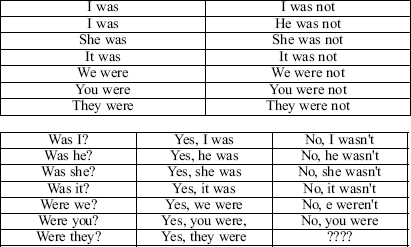
Переведите на английский язык, используя таблицу 6.
1. Я был учеником.
2. Он был летчиком.
3. Она была доктором.
4. Мы были школьниками.
5. Они были рабочими.
6. Ты был рабочим.
7. Они были учениками.
8. Я был дома.
9. Он был в школе
10. Она была в кино?
11. Мы были в парке.
12. Они были в театре?
13. Она была молодая в то время?
14. Он был старый.
15. Она не была учительницей.
16. Они были сильные.
17. Она была больна.
18. Вы были больны?
19. Он был болен?
20. Я не был болен.
21. Я был болен вчера.
22. Она не была больна.
23. Мы были в кино.
24. Они не были в кино.
25. Они не были в школе.
26. Они были дома.
27. Вы были в парке вчера?
28. Он был в школе вчера?
29. Он был рабочим.
30. Она была актрисой.
Answer the questions.
1. What is the pharynx?
2. What shares the pharynx?
3. What has the pharynx?
4. What forms the anterior wall of the laryngopharynx?
5. What the pharyngeal wall con sists of?
6. What is nasopharynx?
7. Where is nasopharynx located?
8. What is the torus tubarius?
9. What is located directly above and behind the torus tubarius?
10. What is located between the pillars that houses the palatine tonsil?
Make the sentences of your own using the new words (10 sentences). Find the verb to be in the text. Explain why it is used in such a way?
Более 800 000 книг и аудиокниг! 📚
Получи 2 месяца Литрес Подписки в подарок и наслаждайся неограниченным чтением
ПОЛУЧИТЬ ПОДАРОКЧитайте также
25-я ЛЕКЦИЯ
25-я ЛЕКЦИЯ Sanguinaria и Chelidonium Прежде чем приступить к изучению Sanguinaria я хочу сказать вам, что есть разновидность мака, растущая в Мексике и называемая Argemone Mexicana. Это растение употребляется в Мексике совершенно таким же образом, как у нас Opium. Оно вызывает также кожные сыпи и
26-я ЛЕКЦИЯ
26-я ЛЕКЦИЯ Cucurbitaceae — Тыквенные Cucurbitaceae:1. Colocyinths cucumis.2. Bryonia alba3. Citrullus. Семена мочегонны.4. Cucurbita (скваш-s quash тыква).5. Momordica balsamum. Ветры.6. Flaterium momordica. Кишки и лихорадка.Сегодня мы начнем изучение Cucurbitaceae. Это семейство дает нам около шести или восьми лекарств, а также несколько
28-я ЛЕКЦИЯ
28-я ЛЕКЦИЯ Coniferae и Euphorbiaceae Coniferae — Хвойные Pinus sylvestris. Детская атрофия. Abies nigra. Желудок. Sabina Juniperus. Выкидыш.Terebenthina.1. Почки, мочевой пузырь и пр.2. Слизистые оболочки.3. Матка.4. Тифозные состояния.5. Почечная водянка.Сравните — Arsenicum, Cantharis, Copaiva, Camphora, Phosphorus.Pix liquida. Легкие
29-я ЛЕКЦИЯ
29-я ЛЕКЦИЯ Ranunculaceae — Лютиковые Aconitum.Helleborus niger.Paeonia.Pulsatilla.Hydrastis.Staphisagria.Actea racemosa.Actea spicata.Radix coptidis.Ranunculus
33-я ЛЕКЦИЯ
33-я ЛЕКЦИЯ Rubiaceae — Мареновые Rubiaceae:1. Rubia titctoiria (Марена).2. Galium (Тоже красная краска).3. Cinchona.4. Ipecacuanha.5. Coffea.6. Mitchella.7. Gambier.Сегодня перед нами семейство растений, из которого мы получаем три очень ценных средства, Cinchona, Ipecacuanha и Coffea. Это семейство дает нам также Gambier (Gambogia,
35-я ЛЕКЦИЯ
35-я ЛЕКЦИЯ Scrophulariaceae — Норичниковые China. Из этого семейства растений мы получаем Digitalis, Gratiola, Leptandra viginica, Euphrasia, Verbascum и Linaria. У нас имеется немного симптомов для каждого из этих средств, и те, которые известны, достаточно определенны, чтобы их легко запомнить. Важнейшим
37-я ЛЕКЦИЯ
37-я ЛЕКЦИЯ Solanaceae — Пасленовые Solanaceae:1. Belladonna.2. Hyoscyamus.3. Stramonium.4. Solan um nigr.5. Tabacum.6. Dulcamara.7. Capsicum.Средства, образующие эту группу по своей симптоматологии очень сходны друг с другом. Едва ли найдется хоть один симптом у этих средств, который не встречался бы почти в том же виде
42-я ЛЕКЦИЯ
42-я ЛЕКЦИЯ Минеральная группа В прилагаемой таблице я разместил для вашего изучения элементы по их взаимному соотношению до некоторой степени так же, как мы находим это в химии. Поэтому они не расположены в порядке, принятом в фармакологии. Но ведь это не абсолютный
44-я ЛЕКЦИЯ
44-я ЛЕКЦИЯ Угольная группа 1. Carbo animalis (содержит фосфат извести).2. Carbo vegetabilis (содержит углекислое кали).3. Graphites (содержит железо).4. Anilin-sulphat.5. Carboneum (сажа).6. Угольный газ.7. Бисульфид угля (сероуглерод).Сегодня я займу ваше внимание лекарствами, получаемыми из угольной
47-я ЛЕКЦИЯ
47-я ЛЕКЦИЯ Acida (Кислоты) Ac. fluoricum, Плавиковая кислотаAc. muriaticum, Соляная кислотаAc. nitricum, Азотная кислотаAc. sulphuricum, Серная кислотаAc. oxalicum, Щавелевая кислотаAc. citricum, Лимонная кислотаAc. phosphoricum, Фосфорная кислотаAc. hudrocuanicum, Синеродисто-водородная кислотаAc. picricum, Пикриновая кислотаAc.
62-я ЛЕКЦИЯ
62-я ЛЕКЦИЯ Препараты Аммония Сегодня у нас на очереди соли Аммония. На таблице вам приведены несколько солей его: Углекислый аммоний, Ammonium carb., Едкий аммиак, Ammonium causticum, Солянокислый аммоний, Ammonium muriaticum, и Фосфорнокислый аммоний, Ammonium phosphoricum. Соли Аммония, взятые как
63-я ЛЕКЦИЯ
63-я ЛЕКЦИЯ Соли извести (Calcarea) Существует целый ряд солей извести, более или менее хорошо исследованных. Первая на этой таблице стоит Calcarea ostrearum или устричная известь. Этот препарат, данный нам Ганеманом, был испытан в качестве углекислой соли извести, Calcarea carbonica. Его
40. Pharynx and related areas
40. Pharynx and related areas The pharynx is a passageway shared by the digestive and respira tory systems. It has lateral, posterior, and medial walls through out, but is open interiorly in its upper regions, communicating with the nasal cavity and the oral cavity. The anterior wall of the laryngopharynx is formed by the larynx. The pharyngeal wall con sists of a mucosa, a fibrous layer, and a muscularis, which is com posed of an inner longitudinal layer and an outer circular
ЛЕКЦИЯ № 1. Вводная лекция. Медицинская символика различных времен и народов
ЛЕКЦИЯ № 1. Вводная лекция. Медицинская символика различных времен и народов История медицины – это наука о развитии, совершенствовании медицинских знаний, медицинской деятельности разных народов мира на протяжении всей истории человечества, которая находится в
Лекция 1
Лекция 1 Еще раз здравствуйте, уважаемые соратники.Начинаем наше первое занятие в Народном университете здорового образа жизни по методу Геннадия Андреевича Шичко. Курс будет посвящен коррекции зрения по методу Шичко-Бейтса. Это курс общего оздоровления и избавления от
Лекция 2
Лекция 2 Итак, здравствуйте, уважаемые соратники.Начинаем наше второе занятие в Народном университете здорового образа жизни по методу Геннадия Андреевича Шичко. Курс посвящен коррекции зрения по методу Шичко-Бейтса. Это курс общего оздоровления и избавления от вредных